Veterinary Diagnostic Tools: Latest Technologies
Microscopy Techniques
Microscopy plays a crucial role in histopathology, enabling the visualization and analysis of tissue samples at various magnifications. Different types of microscopes, including bright-field, fluorescence, and confocal microscopes, are employed for specific applications. These techniques provide detailed images of cellular structures and allow for the identification of various cellular components, such as nuclei, cytoplasm, and organelles. Proper microscopy techniques are essential for accurate and reliable diagnoses in histopathology. Using these techniques, pathologists can gain valuable insights into the health and disease status of tissues.
The selection of the appropriate microscopy technique depends on the specific research question or diagnostic need. For instance, bright-field microscopy is a fundamental technique for routine observations, while fluorescence microscopy is useful for detecting specific molecules or structures within the tissue. Confocal microscopy provides high-resolution images with minimal blurring, offering a powerful tool for detailed three-dimensional analysis.
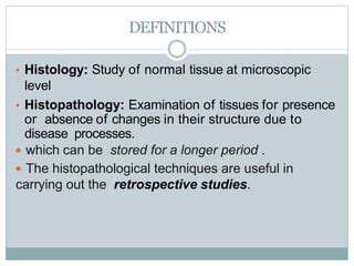
Histopathological Analysis
Histopathological analysis is a crucial step in understanding the characteristics of tissues and cells. This involves examining tissue sections under a microscope to identify structural changes indicative of disease. The examination process involves meticulous observation of tissue architecture, cellular morphology, and the presence of any abnormal structures or cellular components. This careful analysis allows pathologists to diagnose various diseases and conditions.
Pathologists use a standardized approach to evaluate tissue samples, including specific staining techniques that enhance the visibility of different cellular components. Hematoxylin and eosin (H&E) staining is commonly used to highlight nuclei and cytoplasm, providing a basis for general tissue assessment. Other special stains can be used to highlight specific components, such as specific proteins or microorganisms.
Specimen Preparation and Staining
Proper specimen preparation is fundamental to successful histopathological analysis. This involves carefully obtaining the tissue sample, fixing it to preserve its structure, embedding it in a suitable medium, and then sectioning it into thin slices. These meticulous steps ensure that the tissue structure is preserved during the process and allows for clear visualization of the cells and tissues under the microscope. The quality of the specimen preparation directly impacts the reliability of the diagnostic results.
Staining techniques are critical for improving contrast and highlighting specific cellular components. Different staining protocols are employed to highlight particular structures or cellular components, enabling pathologists to identify subtle changes in tissue morphology associated with various diseases. The choice of staining method is often guided by the specific clinical question or the suspected pathology.
Emerging Technologies: Exploring the Frontiers
Advanced Imaging Techniques
Cutting-edge imaging technologies are revolutionizing veterinary diagnostics. High-resolution ultrasound, for example, allows for detailed visualization of internal structures, providing valuable insights into organ function and detecting subtle abnormalities that might be missed with traditional methods. Veterinarians can use this technology to assess the health of organs like the heart, liver, and kidneys, enabling early detection of diseases and tailoring treatment plans accordingly. Furthermore, advancements in magnetic resonance imaging (MRI) are making this powerful modality accessible for use in veterinary medicine, providing exceptional soft tissue contrast and allowing for more precise diagnoses, especially in cases involving the nervous system.
Specialized imaging techniques, such as contrast-enhanced ultrasound, are also emerging, allowing for a more nuanced understanding of blood flow within organs. This enhanced visualization capabilities are crucial for diagnosing vascular diseases, tumors, and inflammatory conditions. The combined use of various imaging modalities offers a comprehensive approach to diagnosis, ensuring a more thorough and accurate assessment of a patient's condition.
Molecular Diagnostics
Molecular diagnostics are rapidly transforming veterinary medicine, enabling the identification of specific genetic mutations and infectious agents. This technology allows for the detection of pathogens at an earlier stage than traditional methods. For instance, polymerase chain reaction (PCR) tests can pinpoint the presence of viruses, bacteria, or parasites, even in very low concentrations, leading to earlier and more effective interventions.
Genetic testing is becoming increasingly important in veterinary medicine. By analyzing an animal's DNA, veterinarians can identify genetic predispositions to certain diseases. This information is invaluable for preventative care, allowing for targeted interventions and potentially preventing the development of debilitating conditions. Early detection of genetic predispositions can lead to proactive management strategies and improved animal health outcomes.
Artificial Intelligence in Diagnostics
Artificial intelligence (AI) is rapidly finding its way into veterinary diagnostics, offering powerful tools for analyzing complex data and assisting in the diagnosis process. AI algorithms can be trained on vast amounts of medical images and patient data to identify patterns and anomalies that might be missed by human observers. This technology has the potential to improve diagnostic accuracy and speed, leading to faster and more effective treatment decisions.
Machine learning models can analyze radiographic images, such as X-rays and CT scans, to identify potential fractures, tumors, or other abnormalities. Furthermore, AI-powered systems can analyze clinical data, such as blood test results and medical history, to predict the likelihood of certain diseases and guide treatment plans. This integration of AI tools into veterinary practices promises to significantly enhance diagnostic capabilities and improve patient outcomes.
Point-of-Care Testing
Point-of-care testing (POCT) provides rapid diagnostic results directly at the animal's bedside, eliminating the need for transporting samples to a laboratory. This technology is invaluable in emergency situations or for animals that are unable to travel. Rapid diagnostic tests for various conditions, including infectious diseases, are becoming increasingly accessible and affordable, empowering veterinarians to make immediate treatment decisions.
Many POCT devices provide results within minutes, enabling veterinarians to initiate appropriate treatment protocols promptly, particularly in critical cases. This immediate feedback loop is crucial for optimizing treatment strategies and improving the chances of a positive outcome. The availability of POCT is transforming how veterinary care is delivered, especially in remote or underserved areas.
Remote Monitoring and Telemedicine
Remote monitoring technologies are transforming how veterinarians manage the health of animals, allowing for continuous tracking of vital signs and behavior. Wearable sensors and other remote monitoring devices can transmit data to veterinary clinicians, providing real-time insights into the patient's condition, enabling proactive interventions and reducing the need for frequent in-person visits. This is particularly helpful for animals with chronic conditions or those that are geographically isolated.
Telemedicine platforms are bridging the gap between veterinarians and their clients, enabling remote consultations and advice. This technology allows veterinarians to provide guidance and support to clients, especially in cases of minor illnesses or when in-person visits are not immediately possible. Remote monitoring and telemedicine are fostering a more accessible and responsive approach to veterinary care, improving the overall well-being of animals.
Nanotechnology in Diagnostics
Nanotechnology is poised to revolutionize veterinary diagnostics, offering highly targeted and sensitive tools for detecting diseases at the molecular level. Nanoparticles can be designed to specifically bind to disease markers, enabling earlier detection and more precise diagnosis. This technology holds immense promise for identifying pathogens, monitoring drug delivery, and assessing tissue damage with greater accuracy.
The development of nanomaterials for targeted drug delivery is another exciting application. Using nanoparticles, drugs can be delivered directly to affected areas, minimizing side effects and maximizing therapeutic efficacy. This precision approach is particularly valuable in treating complex diseases and improving treatment outcomes for animals.
Read more about Veterinary Diagnostic Tools: Latest Technologies


Hot Recommendations
- Best Pet Bowls: Stainless Steel and Ceramic
- Pet Hydration: Why It's Crucial
- Stop Counter Surfing: Training Your Dog to Stay Off
- Pet Hypothyroidism: Symptoms and Management
- Signs of Pet Liver Disease: What to Watch For
- Pet Emergency Kits: What to Pack
- Dangers of Xylitol: Toxic to Dogs
- Dealing with Pet Diarrhea: When to See a Vet
- Preparing Pets for Travel: Tips for a Smooth Trip
- Pet Depression: Recognizing the Signs